Toxoplasma gondii (/ˈtɒksoʊplæzmə ˈɡɒndiaɪ/) is an obligate intracellular parasitic protozoan (specifically an apicomplexan) that causes toxoplasmosis.[3] Found worldwide, T. gondii is capable of infecting virtually all warm-blooded animals,[4]: 1 but felids, such as domestic cats, are the only known definitive hosts in which the parasite may undergo sexual reproduction.[5][6]
T. gondii has been shown to alter the behavior of infected rodents in ways that increase the rodents' chances of being preyed upon by felids.[7][8][9] Support for this "manipulation hypothesis" stems from studies showing that T. gondii-infected rats have a decreased aversion to cat urine.[7] Because cats are the only hosts within which T. gondii can sexually reproduce to complete and begin its lifecycle, such behavioral manipulations are thought to be evolutionary adaptations that increase the parasite's reproductive success.[7] Rats that do not avoid cats' habitations will more likely become cat prey.
Toxoplasma gondii infection in mice lowers general anxiety, increases explorative behaviors and surprisingly increases a general loss of aversion to predators without selectivity toward cats. There is a positive correlation between the severity of the behavioral alterations and the cyst load, which reflects indirectly the level of inflammation during brain colonization. These results point toward unspecific and immune related changes in the infected mice brains associated with altered behaviors. These results refute the thought of a selective loss of cat fear with possible reference to favoring non-sexual transmission between intermediate hosts.[10]
The primary mechanisms of T. gondii–induced behavioral changes in rodents are now known to occur through epigenetic remodeling in neurons that govern the relevant behaviors;[11][12] which, for example, modifies epigenetic methylation to cause hypomethylation of arginine vasopressin-related genes in the medial amygdala, such that predator aversion is greatly decreased.[11][12] Widespread histone-lysine acetylation in cortical astrocytes appears to be another epigenetic mechanism employed by T. gondii.[13][failed verification][14]
Differences are also observed between infected and non-infected humans in their aversion to cat urine, but with divergent trajectories by gender.[15]
In humans, particularly infants and those with weakened immunity, such as HIV/AIDS patients; T. gondii infection may cause a serious – and occasionally fatal – illness: toxoplasmosis.[16][4]: 77
T. gondii is one of the most common parasites in developed countries;[17][18] serological studies estimate that 30–50% of the global population has been exposed to, and may be chronically infected with, T. gondii; although infection rates differ significantly from country to country.[19][20] For example, estimates have shown the highest IgG seroprevalence to be in Ethiopia, at 64.2%, as of 2018.[21]
Mild, flu-like symptoms occasionally occur during the first few weeks following exposure; otherwise, symptoms are not readily observable in healthy human adults.[19][16][4]: 77 This asymptomatic state of infection is referred to as a latent infection, and it has recently been associated with numerous subtle, yet adverse or pathological, behavioral alterations in humans,[19][22] though some newer studies found this association to be weak and concluded:
"On the whole, there was little evidence that T. gondii was related to increased risk of psychiatric disorder, poor impulse control, personality aberrations or neurocognitive impairment." [23]
A number of studies have suggested that subtle behavioral or personality changes may occur in infected humans,[24] and infection with the parasite has recently been associated with a number of neurological disorders – particularly schizophrenia[9] and bipolar disorder.[25][26] A 2015 study also found cognitive deficits in adults to be associated with joint infection by both T. gondii and Helicobacter pylori in a regression model with controls for race-ethnicity and educational attainment.[27] Although a causal relationship between latent toxoplasmosis with these neurological phenomena has not yet been established,[19][9] preliminary evidence suggests that T. gondii infection may induce some of the same alterations in the human brain as those observed in mice.[28][29]
Structure

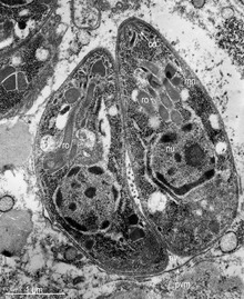
T. gondii contains organelles called rhoptries and micronemes, as well as other organelles.
Lifecycle

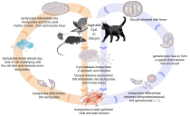
The lifecycle of T. gondii may be broadly summarized into two components: a sexual component that occurs only within cats (felids, wild or domestic), and an asexual component that can occur within virtually all warm-blooded animals, including humans, cats, and birds.[30]: 2 Because T. gondii can sexually reproduce only within cats, cats are therefore the definitive host of T. gondii. All other hosts – in which only asexual reproduction can occur – are intermediate hosts.
Sexual reproduction in the feline definitive host
When a feline is infected with T. gondii (e.g. by consuming an infected mouse carrying the parasite's tissue cysts), the parasite survives passage through the stomach, eventually infecting epithelial cells of the cat's small intestine.[30]: 39 Inside these intestinal cells, the parasites undergo sexual development and reproduction, producing millions of thick-walled, zygote-containing cysts known as oocysts. Felines are the only definitive host because they lack expression of the enzyme delta-6-desaturase (D6D) in their intestine. This enzyme converts linoleic acid; the absence of expression allows systemic linoleic acid accumulation. Recent findings showed that this excess of linoleic acid is essential for T. gondii sexual reproduction.[6]

Feline shedding of oocysts
Infected epithelial cells eventually rupture and release oocysts into the intestinal lumen, whereupon they are shed in the cat's feces.[4]: 22 Oocysts can then spread to soil, water, food, or anything potentially contaminated with the feces. Highly resilient, oocysts can survive and remain infective for many months in cold and dry climates.[31]
Ingestion of oocysts by humans or other warm-blooded animals is one of the common routes of infection.[32] Humans can be exposed to oocysts by, for example, consuming unwashed vegetables or contaminated water, or by handling the feces (litter) of an infected cat.[30]: 2 [33] Although cats can also be infected by ingesting oocysts, they are much less sensitive to oocyst infection than are intermediate hosts.[34][4]: 107
Initial infection of the intermediate host
Intermediate hosts found include pigs, chickens, goats, sheep[30]: 2 and Macropus rufus by Moré et al 2010.[35]: 162 Cattle and horses are resistant and thought to be incapable of significant infection.[30]: 11 T. gondii is considered to have three stages of infection; the tachyzoite stage of rapid division, the bradyzoite stage of slow division within tissue cysts, and the oocyst environmental stage.[36] Tachyzoites are also known as "tachyzoic merozoites" and bradyzoites as "bradyzoic merozoites".[37] When an oocyst or tissue cyst is ingested by a human or other warm-blooded animal, the resilient cyst wall is dissolved by proteolytic enzymes in the stomach and small intestine, freeing sporozoites from within the oocyst.[32][36] The parasites first invade cells in and surrounding the intestinal epithelium, and inside these cells, the parasites differentiate into tachyzoites, the motile and quickly multiplying cellular stage of T. gondii.[30]: 39 Tissue cysts in tissues such as brain and muscle tissue, form about 7–10 days after initial infection.[36] Although severe infection of M. rufus has been observed it is unknown whether this is common.[35]
Asexual reproduction in the intermediate host
Inside host cells, the tachyzoites replicate inside specialized vacuoles (called the parasitophorous vacuoles) created from host cell membrane during invasion into the cell.[30]: 23–39 Tachyzoites multiply inside this vacuole until the host cell dies and ruptures, releasing and spreading the tachyzoites via the bloodstream to all organs and tissues of the body, including the brain.[30]: 39–40
Growth in tissue culture
The parasite can be easily grown in monolayers of mammalian cells maintained in vitro in tissue culture. It readily invades and multiplies in a wide variety of fibroblast and monocyte cell lines. In infected cultures, the parasite rapidly multiplies and thousands of tachyzoites break out of infected cells and enter adjacent cells, destroying the monolayer in due course. New monolayers can then be infected using a drop of this infected culture fluid and the parasite indefinitely maintained without the need of animals.

Formation of tissue cysts
Following the initial period of infection characterized by tachyzoite proliferation throughout the body, pressure from the host's immune system causes T. gondii tachyzoites to convert into bradyzoites, the semidormant, slowly dividing cellular stage of the parasite.[38] Inside host cells, clusters of these bradyzoites are known as tissue cysts. The cyst wall is formed by the parasitophorous vacuole membrane.[30]: 343 Although bradyzoite-containing tissue cysts can form in virtually any organ, tissue cysts predominantly form and persist in the brain, the eyes, and striated muscle (including the heart).[30]: 343 However, specific tissue tropisms can vary between intermediate host species; in pigs, the majority of tissue cysts are found in muscle tissue, whereas in mice, the majority of cysts are found in the brain.[30]: 41
Cysts usually range in size between five and 50 µm in diameter,[39] (with 50 µm being about two-thirds the width of the average human hair).[40]
Consumption of tissue cysts in meat is one of the primary means of T. gondii infection, both for humans and for meat-eating, warm-blooded animals.[30]: 3 Humans consume tissue cysts when eating raw or undercooked meat (particularly pork and lamb).[41] Tissue cyst consumption is also the primary means by which cats are infected.[4]: 46
An exhibit at the San Diego Natural History Museum states urban runoff with cat feces transports Toxoplasma gondii into the ocean, which can kill sea otters.[42]
Chronic infection
Tissue cysts can be maintained in host tissue for the lifetime of the animal.[30]: 580 However, the perpetual presence of cysts appears to be due to a periodic process of cyst rupturing and re-encysting, rather than a perpetual lifespan of individual cysts or bradyzoites.[30]: 580 At any given time in a chronically infected host, a very small percentage of cysts are rupturing,[30]: 45 although the exact cause of this tissue cysts rupture is, as of 2010, not yet known.[4]: 47
Theoretically, T. gondii can be passed between intermediate hosts indefinitely via a cycle of consumption of tissue cysts in meat. However, the parasite's lifecycle begins and completes only when the parasite is passed to a feline host, the only host within which the parasite can again undergo sexual development and reproduction.[32]
Population structure in the wild
Khan et al.[43] reviewed evidence that despite the occurrence of a sexual phase in its life cycle, T. gondii has an unusual population structure dominated by three clonal lineages (Types I, II and III) that occur in North America and Europe. They estimated that a common ancestor founded these about 10,000 years ago. In a further and larger study (with 196 isolates from diverse sources including T. gondii found in the bald eagle, gray wolves, Arctic foxes and sea otters), Dubey et al.[44] also found that T. gondii strains infecting North American wildlife have limited genetic diversity with the occurrence of only a few major clonal types. They found that 85% of strains in North America were of one of three widespread genotypes (Types II, III and Type 12). Thus T. gondii has retained the capability for sex in North America over many generations, producing largely clonal populations, and matings have generated little genetic diversity.
Cellular stages
During different periods of its life cycle, individual parasites convert into various cellular stages, with each stage characterized by a distinct cellular morphology, biochemistry, and behavior. These stages include the tachyzoites, merozoites, bradyzoites (found in tissue cysts), and sporozoites (found in oocysts).
Some stages are motile and some calcium-dependent protein kinases (TgCDPKs) are involved in this parasite's motility.[45][46] Gaji et al 2015 find TgCDPK3 is required to begin the action of motility because it phosphorylates T. gondii's myosin A (TgMYOA).[45][46] TgCDPK3 is the functional orthologue of CDPK1 in this parasite.[46]
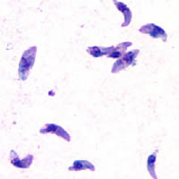
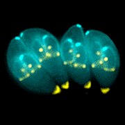
Tachyzoites
Motile, and quickly multiplying, tachyzoites are responsible for expanding the population of the parasite in the host.[47][30]: 19 When a host consumes a tissue cyst (containing bradyzoites) or an oocyst (containing sporozoites), the bradyzoites or sporozoites stage-convert into tachyzoites upon infecting the intestinal epithelium of the host.[30]: 359 During the initial acute period of infection, tachyzoites spread throughout the body via the blood stream.[30]: 39–40 During the later, latent (chronic) stages of infection, tachyzoites stage-convert to bradyzoites to form tissue cysts.
Merozoites

Like tachyzoites, merozoites divide quickly, and are responsible for expanding the population of the parasite inside the cat's intestine before sexual reproduction.[30]: 19 When a feline definitive host consumes a tissue cyst (containing bradyzoites), bradyzoites convert into merozoites inside intestinal epithelial cells. Following a brief period of rapid population growth in the intestinal epithelium, merozoites convert into the noninfectious sexual stages of the parasite to undergo sexual reproduction, eventually resulting in zygote-containing oocysts.[30]: 306
Bradyzoites
Bradyzoites are the slowly dividing stage of the parasite that make up tissue cysts. When an uninfected host consumes a tissue cyst, bradyzoites released from the cyst infect intestinal epithelial cells before converting to the proliferative tachyzoite stage.[30]: 359 Following the initial period of proliferation throughout the host body, tachyzoites then convert back to bradyzoites, which reproduce inside host cells to form tissue cysts in the new host.
Sporozoites
Sporozoites are the stage of the parasite residing within oocysts. When a human or other warm-blooded host consumes an oocyst, sporozoites are released from it, infecting epithelial cells before converting to the proliferative tachyzoite stage.[30]: 359
Immune response
Initially, a T. gondii infection stimulates production of IL-2 and IFN-γ by the innate immune system.[38] Continuous IFN-γ production is necessary for control of both acute and chronic T. gondii infection.[38] These two cytokines elicit a CD4+ and CD8+ T-cell mediated immune response.[38] Thus, T-cells play a central role in immunity against Toxoplasma infection. T-cells recognize Toxoplasma antigens that are presented to them by the body's own Major Histocompatibility Complex (MHC) molecules. The specific genetic sequence of a given MHC molecule differs dramatically between individuals, which is why these molecules are involved in transplant rejection. Individuals carrying certain genetic sequences of MHC molecules are much more likely to be infected with Toxoplasma. One study of >1600 individuals found that Toxoplasma infection was especially common among people who expressed certain MHC alleles (HLA-B*08:01, HLA-C*04:01, HLA-DRB 03:01, HLA-DQA*05:01 and HLA-DQB*02:01).[48]
IL-12 is produced during T. gondii infection to activate natural killer (NK) cells.[38] Tryptophan is an essential amino acid for T. gondii, which it scavenges from host cells. IFN-γ induces the activation of indole-amine-2,3-dioxygenase (IDO) and tryptophan-2,3-dioxygenase (TDO), two enzymes that are responsible for the degradation of tryptophan.[49] Immune pressure eventually leads the parasite to form cysts that normally are deposited in the muscles and in the brain of the hosts.[38]
Immune response and behavior alterations
The IFN-γ-mediated activation of IDO and TDO is an evolutionary mechanism that serves to starve the parasite, but it can result in depletion of tryptophan in the brain of the host. IDO and TDO degrade tryptophan to N-formylkynurenine. Administration of L-kynurenine is capable of inducing depressive-like behavior in mice.[49] T. gondii infection has been demonstrated to increase the levels of kynurenic acid (KYNA) in the brains of infected mice and KYNA has also been demonstrated to be increased in the brain of schizophrenic persons.[49] Low levels of tryptophan and serotonin in the brain were already associated with depression.[50]
Risk factors for human infection
The following have been identified as being risk factors for T. gondii infection in humans and warm-blooded animals:
- by consuming raw or undercooked meat containing T. gondii tissue cysts.[33][51][52][53][54] The most common threat to citizens in the United States is from eating raw or undercooked pork.[55]
- by ingesting water, soil, vegetables, or anything contaminated with oocysts shed in the feces of an infected animal.[51] Cat fecal matter is particularly dangerous: Just one cyst consumed by a cat can result in thousands of oocysts. This is why physicians recommend pregnant or ill persons do not clean the cat's litter box at home.[55] These oocysts are resilient to harsh environmental conditions and can survive over a year in contaminated soil.[36][56]
- from a blood transfusion or organ transplant[57]
- from transplacental transmission from mother to fetus, particularly when T. gondii is contracted during pregnancy[51]
- from drinking unpasteurized goat milk[52]
- from raw and treated sewage and bivalve shellfish contaminated by treated sewage[58][59][60][61]
A common argument in the debate about whether cat ownership is ethical involves the question of Toxoplasma gondii transmission to humans.[62] Even though "living in a household with a cat that used a litter box was strongly associated with infection,"[33] and that living with several kittens or any cat under one year of age has some significance,[63] several other studies claim to have shown that living in a household with a cat is not a significant risk factor for T. gondii infection. [53][64]
Specific vectors for transmission may also differ based on geographic location. "The seawater in California is thought to be contaminated by T. gondii oocysts that originate from cat feces, survive or bypass sewage treatment, and travel to the coast through river systems. T. gondii has been identified in a California mussel by polymerase chain reaction and DNA sequencing. In light of the potential presence of T. gondii, pregnant women and immunosuppressed persons should be aware of this potential risk associated with eating raw oysters, mussels, and clams.[52]
In warm-blooded animals, such as brown rats, sheep, and dogs, T. gondii has also been shown to be sexually transmitted.[65][66][67] Although T. gondii can infect, be transmitted by, and asexually reproduce within humans and virtually all other warm-blooded animals, the parasite can sexually reproduce only within the intestines of members of the cat family (felids).[32] Felids are therefore the definitive hosts of T. gondii; all other hosts (like human or other mammals) are intermediate hosts.
Preventing infection
The following precautions are recommended to prevent or greatly reduce the chances of becoming infected with T. gondii. This information has been adapted from the websites of United States Centers for Disease Control and Prevention[68] and the Mayo Clinic.[69]
From food
Basic food-handling safety practices can prevent or reduce the chances of becoming infected with T. gondii, such as washing unwashed fruits and vegetables, and avoiding raw or undercooked meat, poultry, and seafood. Other unsafe practices such as drinking unpasteurized milk or untreated water can increase odds of infection.[68] As T. gondii is commonly transmitted through ingesting microscopic cysts in the tissues of infected animals, meat that is not prepared to destroy these presents a risk of infection. Freezing meat for several days at subzero temperatures (0 °F or −18 °C) before cooking may break down all cysts, as they rarely survive these temperatures.[4]: 45 During cooking, whole cuts of red meat should be cooked to an internal temperature of at least 145 °F (63 °C). Medium rare meat is generally cooked between 130 and 140 °F (55 and 60 °C),[70] so cooking meat to at least medium is recommended. After cooking, a rest period of 3 min should be allowed before consumption. However, ground meat should be cooked to an internal temperature of at least 160 °F (71 °C) with no rest period. All poultry should be cooked to an internal temperature of at least 165 °F (74 °C). After cooking, a rest period of 3 min should be allowed before consumption.
From environment
Oocysts in cat feces take at least a day to sporulate (to become infectious after they are shed), so disposing of cat litter daily greatly reduces the chance of infectious oocysts developing. As these can spread and survive in the environment for months, humans should wear gloves when gardening or working with soil, and should wash their hands promptly after disposing of cat litter. These precautions apply to outdoor sandboxes/play sand pits, which should be covered when not in use. Cat feces should never be flushed down a toilet.
Pregnant women are at higher risk of transmitting the parasite to their unborn child and immunocompromised people of acquiring a lingering infection. Because of this, they should not change or handle cat litter boxes. Ideally, cats should be kept indoors and fed only food that has low to no risk of carrying oocysts, such as commercial cat food or well-cooked table food.
Vaccination
As of 2016, no approved human vaccine exists against Toxoplasma gondii.[71] Research on human vaccines is ongoing.[72]
For sheep, an approved live vaccine sold as Toxovax (from MSD Animal Health) provides lifetime protection.[73]
Treatment
In humans, active toxoplasmosis can be treated with a combination of drugs such as pyrimethamine and sulfadiazine, plus folinic acid. Immune-compromised patients may need continuous treatment until/unless their immune system is restored.[74]
Environmental effects
In many parts of the world, where there are high populations of feral cats, there is an increased risk to the native wildlife due to increased infection of Toxoplasma gondii. It has been found that the serum concentrations of T. gondii in the wildlife population were increased where there are high amounts of cat populations. This creates a dangerous environment for organisms that have not evolved in cohabitation with felines and their contributing parasites. [75]
Impact on marine species
Minks and otters
Toxoplasmosis is one of the contributing factors towards mortality in southern sea otters, especially in areas where there is large urban run-off.[76] In their natural habitats, sea otters control sea urchin populations and, thus indirectly, control sea kelp forests. By enabling the growth of sea kelp, other marine populations are protected as well as CO2 emissions are reduced due to the kelp's ability to absorb atmospheric carbon.[77] An examination on 105 beachcast otters revealed that 38.1% had parasitic infections, and 28% of said infections had resulted in protozoal meningoencephalitis deaths.[76] Toxoplasma gondii was found to be the root cause in 16.2% of these deaths, while 6.7% of the deaths were due to a closely related protozoan parasite known as Sarcocystis neurona.[76]
Minks, being semiaquatic, are also susceptible to infection and being antibody-positive toward Toxoplasma gondii.[78] Minks can follow a similar diet as otters and feasts on crustaceans, fish, and invertebrates, thus the transmission route follows a similar pattern to otters. Because of the mink's ability to transverse land more frequently, and often seen as an invasive species itself, minks are a bigger threat in transporting T. gondii to other mammalian species, rather than otters who have a more restrictive breadth.[78]
Black-footed penguins
Although under-studied, penguin populations, especially those that share an environment with the human population, are at-risk due to parasite infections, mainly Toxoplasmosis gondii. The main subspecies of penguins found to be infected by T. gondii include wild Magellanic and Galapagos penguins, as well as blue and African penguins in captivity.[79] In one study, 57 (43.2%) of 132 serum samples of Magellanic penguins were found to have T. gondii. The island that the penguin is located, Magdalena Island, is known to have no cat populations, but a very frequent human population, indicating the possibility of transmission.[79]
Histopathology
Examination of black-footed penguins with toxoplasmosis reveals hepatomegaly, splenomegaly, cranial hemorrhage, and necrotic kidneys (Ploeg, et al., 2011). Alveolar and hepatic tissue presents a high number of immune cells like macrophages containing tachyzoites of T. gondii.[80] Histopathological features in other animals affected with toxoplasmosis had tachyzoites in eye structures like the retina which lead to blindness.[80]
Water transmission
The transmission of oocysts has been unknown, even though there have many documented cases of infection in marine species. Researchers have found that the oocytes of T. gondii can survive in seawater for at least 6 months, with the amount of salt concentration not affecting its lifecycle. There have been no studies on the ability of T. gondii oocysts lifecycle within freshwater environments, though infections are still present. One possible hypothesis of transmission is via amoeba species, particularly Acanthamoeba spp, a species that is found in all water environments (fresh-, brackish, and full-strength seawater). Normally, amoebas’ function as a natural filter, phagocytizing nutrients and bacteria found within the water. Some pathogens have used this to their advantage, however, and evolved to be able to avoid being broken down and, thus, survive encased in the amoeba – this includes Holosporaceae, Pseudomonaceae, Burkholderiacceae, among others.[81] Overall, this aids the pathogen in transportation but, also, protection from drugs and sterilizers that would, otherwise, cause death in the pathogen.[82] Studies have shown that T. gondii oocysts can live within amoebas after being engulfed for at least 14 days without significant obliteration of the parasite.[83] The ability of the microorganism to survive in vitro is dependent on the microorganism itself, but there are a few overarching mechanisms present. T. gondii oocysts have been found to resist an acidic pH and, thus, are protected by the acidification found in endocytic vacuoles and lysosomes.[83] Phagocytosis further increases with the carbohydrate-rich surface membrane located on the amoebae.[84] The pathogen can be released either by lysis of the amoebae or by exocytosis, but this is understudied [85]